
Single Position Lumbar Fusion and Endoscopic Decompression
Doctor Profile
Dr. Arvind Umarani
MS Orthopedics, KMC Manipal, Fellowship trained Spine Surgeon
Spine Surgeon | India
Specialisation: Minimally invasive and endoscopic spine surgery, deformity correction, navigation-guided spine techniques
This case study is drawn from a published case report co authored by Dr. Umarani as first author, alongside Dr. Masato Tanaka and colleagues in the Department of Orthopaedic Surgery, Okayama Rōsai Hospital, Japan. Dr. Umarani contributed to the concept, design, and drafting of the study, working within the spine surgery team at the hospital.
Key Takeaways
Condition: Recurrent low back and left leg pain from combined lumbar instability at L3 to 4 and nerve root compression at L5 to S1, following prior multilevel decompression surgery
Procedure: Simultaneous lumbar interbody fusion with percutaneous pedicle screw fixation, combined with endoscopic assisted nerve decompression, performed in a single lateral position without fluoroscopy
Doctor: Dr. Arvind Umarani, first author, Okayama Rōsai Hospital, Japan
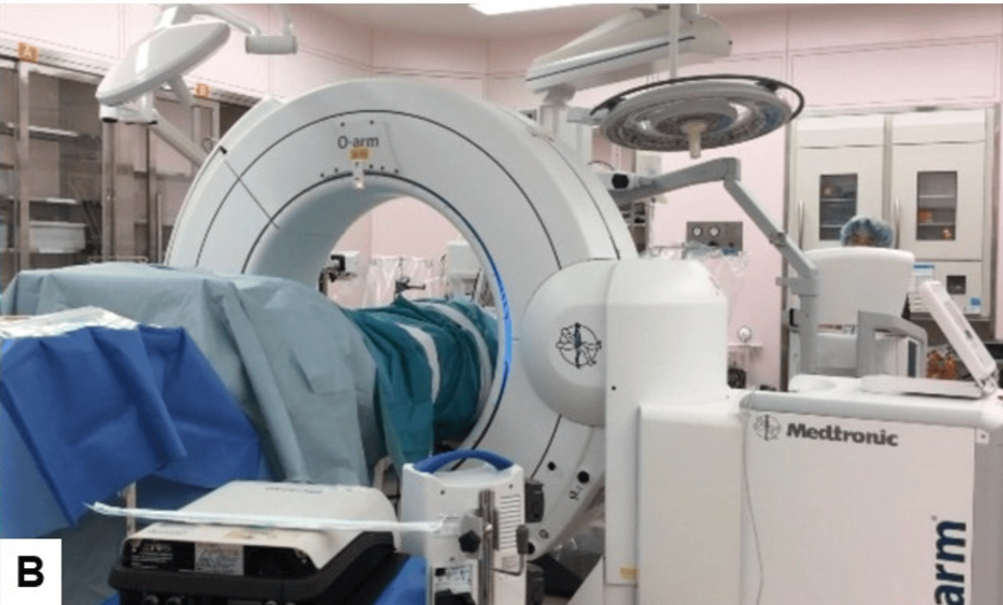
Technique: Navigation guided, fluoroscopy free approach using intraoperative O arm imaging, single position, two surgeon workflow
Outcome: Marked improvement in leg pain and function, with disability score improving from 49 percent to 20 percent at one year follow up
Complications: None. No intraoperative complications and no new neurological deficit
Patient Profile
Field | Details |
Age | 71 years |
Gender | Female |
Relevant history | Rheumatoid arthritis, prior lumbar decompression surgery 3 years earlier |
Presenting complaint | Low back pain and left leg radicular pain, present for 4 years and progressively worsening |
Diagnosis | Recurrent lumbar stenosis with L3 to 4 instability and L5 to S1 foraminal stenosis |
Duration of issue | 4 years, with walking distance reduced to under 200 metres before symptoms limited activity |
Previous treatments | Prior posterior decompression surgery, followed by conservative treatment |
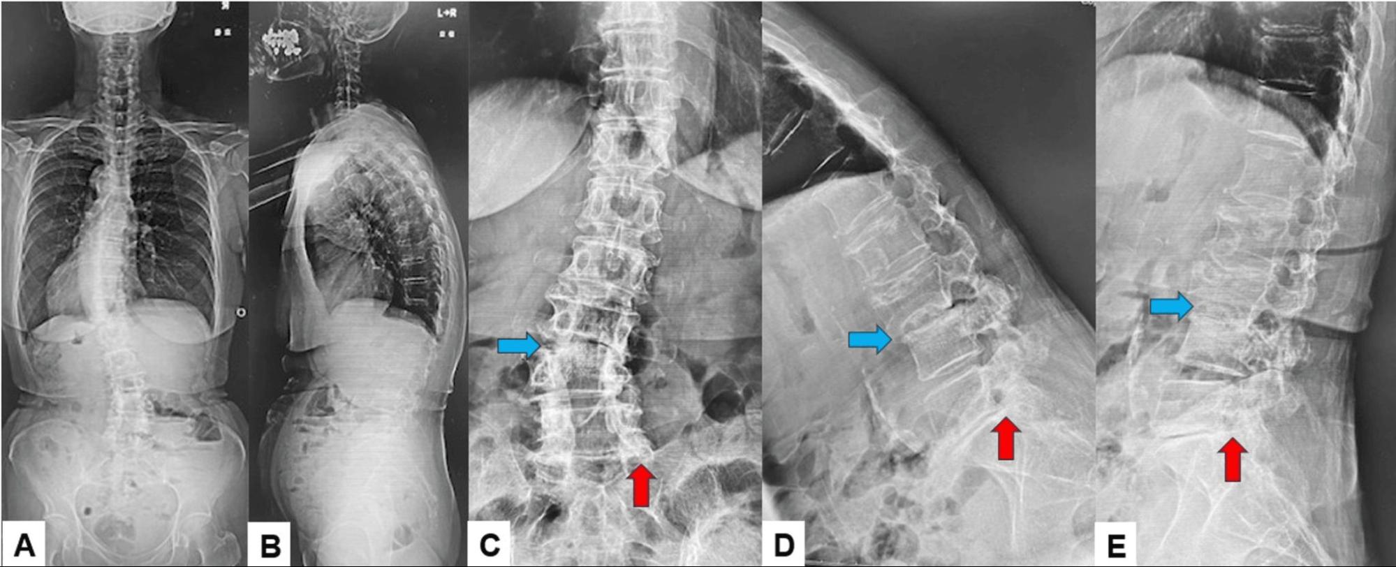
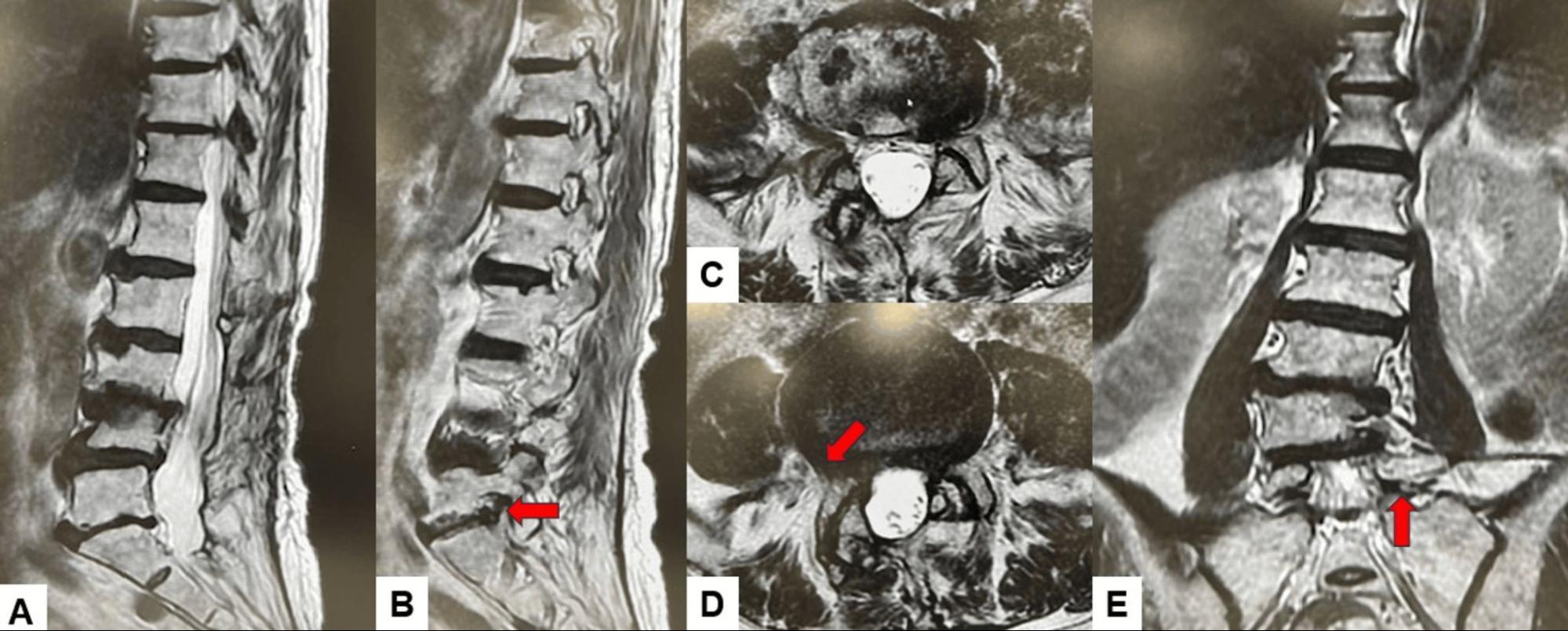
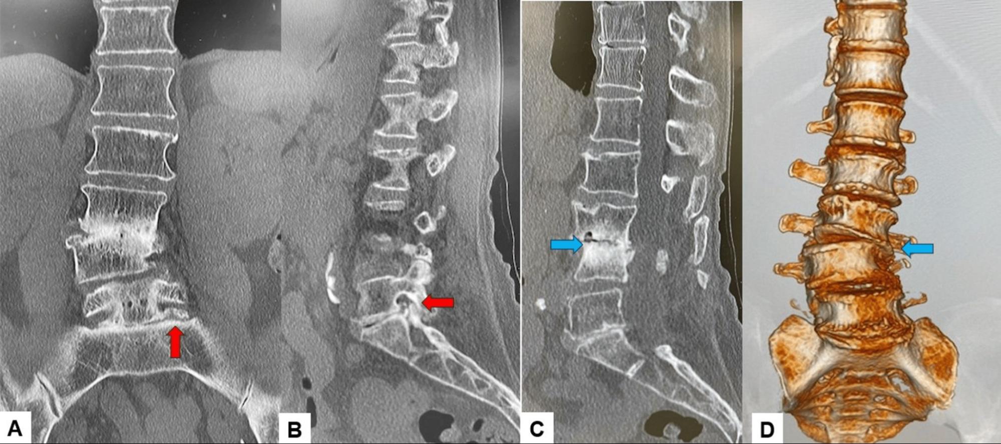
Preoperative findings | L3 to 4 collapse with endplate changes, left L5 to S1 foraminal stenosis, severe compression of the left L5 nerve root |
Date of procedure | Within the published study period, 2026 |
Outcome | Excellent |
Patient identity is withheld in line with confidentiality guidelines and journal publication standards. Data is drawn from a case report published in Cureus, 2026, 18(5): e108565.
The Problem
CONDITION
This patient had two separate but related problems in the lumbar spine. One level, L3 to 4, had become unstable, with the vertebrae shifting slightly out of alignment and the disc space narrowing, which was contributing to persistent low back pain. A second level, L5 to S1, had significant narrowing of the nerve exit canal on the left side, which was compressing the nerve root and causing pain, weakness, and numbness down the left leg. This combination, instability at one level and nerve compression at another, is a common but technically demanding pattern, particularly when a patient has already had prior spinal surgery.
EMOTIONAL AND PSYCHOLOGICAL IMPACT
Living with worsening back and leg pain over several years takes a toll well beyond the physical symptoms. This patient’s walking distance had dropped to under 200 metres before surgery, which meaningfully limits day to day independence and routine activities. Having already undergone one spinal surgery and continued to decline despite conservative treatment can also be discouraging, and understandably raises questions about whether further surgery is worthwhile or safe. For patients in this situation, understanding what modern endoscopic spine surgery can offer, particularly with less tissue disruption than repeat open surgery, is often the most important part of the conversation.
Consultation And Treatment Plan
WHAT WAS ASSESSED DURING CONSULTATION
Walking distance and pattern of leg pain on exertion
Muscle strength and sensation in the affected leg
Standing and flexion extension radiographs to assess spinal alignment and instability
CT imaging to assess bone structure and nerve canal narrowing
MRI to confirm the exact location and severity of nerve compression
The instability at L3 to 4 required fusion, while the nerve compression at L5 to S1 required direct decompression, meaning two different problems needed two different solutions in the same surgery
Performing both procedures in a single lateral position avoided the need to reposition the patient partway through surgery, which reduces operative time and anaesthesia duration
A navigation guided, fluoroscopy free workflow was chosen specifically to eliminate radiation exposure to the patient and surgical team throughout the procedure
This approach was selected in part to avoid additional dissection through previously operated tissue at the level of prior decompression
This decision making reflects a broader principle in modern spine care. An experienced Spine Surgeon increasingly has access to techniques that combine fusion and decompression without repositioning the patient, which can reduce operative time, blood loss, and radiation exposure compared with older staged approaches. An Orthopedic Spine Surgeon evaluating a patient with combined instability and nerve compression will typically weigh which levels need fusion, which need decompression alone, and whether a single position technique is technically appropriate for that patient’s anatomy.
PRE-OPERATIVE IMAGING
Procedure Details
STEP BY STEP OVERVIEW
Patient positioned in the right lateral position on a radiolucent frame, with the table gently flexed to open the disc space
Neuromonitoring leads connected and baseline signals recorded before any surgical step
A percutaneous reference frame placed near the pelvis, followed by an intraoperative scan for surgical navigation, avoiding the need for a C arm throughout the case
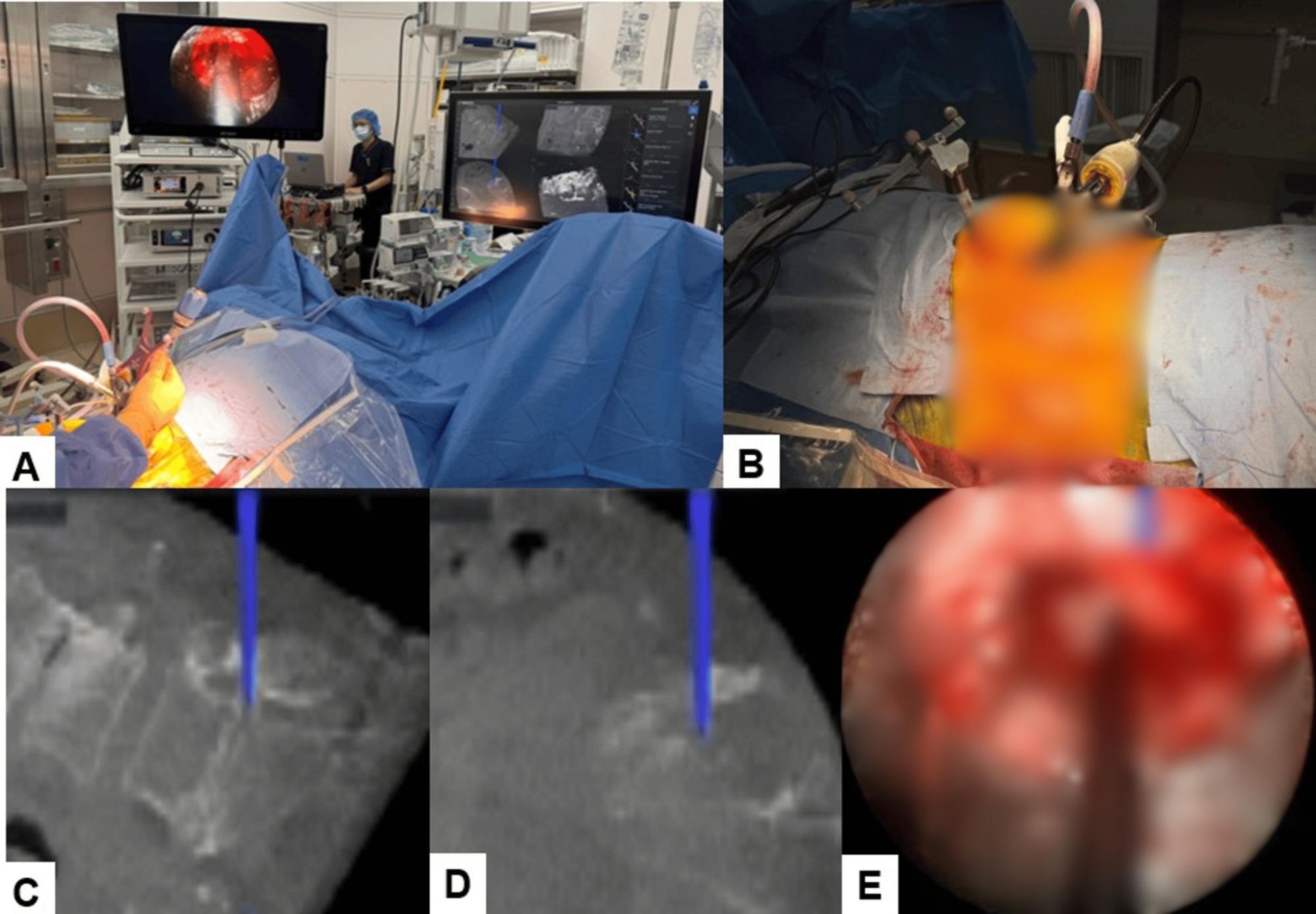
A two surgeon workflow used, with one surgeon performing the lumbar fusion while the second placed the pedicle screws at the same time
A small incision made lateral to the disc space, with gentle retroperitoneal access and careful retraction of the surrounding muscle under direct visualisation
A titanium coated interbody cage packed with bone graft inserted at the unstable level to restore disc height and alignment
Following fusion, the same position used to perform endoscopic decompression at the second level, using a working channel and a high speed endoscopic burr to relieve pressure on the compressed nerve root
No repositioning required at any point during the procedure
Detail | Result |
Approach | Single lateral position, fusion combined with endoscopic decompression |
Anaesthesia | General anaesthesia |
Fluoroscopy used | None. Fully navigation guided |
Operative time | 163 minutes |
Estimated blood loss | 200 mL |
Intraoperative complications | None |
Postoperative mobilisation | Mobilised with a brace on Day 1 |
Post-Operative Results
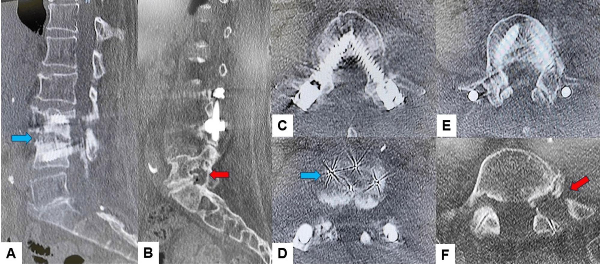
The combined procedure achieved both goals in a single operation, stabilising the unstable level and fully decompressing the affected nerve root, without the need for a second surgery or a change in patient position. Recovery was smooth, with no new neurological problems and steady improvement in both pain and walking distance over the following months.
Outcome Metric | Result |
Operative time | 163 minutes |
Estimated blood loss | 200 mL |
Disability score, before surgery | 49 percent |
Disability score, at 1 year | 20 percent |
Walking distance, at 3 months | Over 1 kilometre without claudication |
Lumbar lordosis | Improved from 25 degrees to 32 degrees |
Foraminal height | Increased from 6.8 mm to 9.4 mm |
Fusion status at 1 year | Progressive fusion, no cage subsidence, no adjacent segment degeneration |
Neurological deficit | None |
Patient Feedback
“After the surgery, my leg pain and numbness improved a lot. I was able to walk better and for a longer distance without pain. I feel much better now and am happy with the improvement.”
Post-Procedure Care And Recovery
INSTRUCTIONS GIVEN TO PATIENTS
Mobilised with a supportive brace from postoperative Day 1
Standard postoperative monitoring for neurological status and wound healing
Scheduled follow up imaging at 3 months and 1 year
RECOVERY TIMELINE
Timeframe | What Patient Can Expect |
Day 1 | Mobilisation begins with a brace, no new neurological deficit observed |
3 months | Walking distance improved to over 1 kilometre without claudication, sensory disturbance nearly resolved |
1 year | Disability score improved from 49 percent to 20 percent, imaging confirmed stable fusion and no complications |
Frequently Asked Questions
Q1. What causes recurrent back and leg pain after previous spine surgery?
Recurrent pain can occur when a different level of the spine becomes unstable or narrowed over time, separate from the level originally treated. In this case, a new level of instability developed above the previous surgery site, alongside nerve compression at another level.
Q2. What is the advantage of performing fusion and decompression without changing patient position?
Avoiding repositioning during surgery reduces operative time, anaesthesia duration, and the risk of contamination from redraping. It also allows two connected problems to be treated in a single, continuous procedure rather than as separate operations.
Q3. Is it possible to perform this kind of spine surgery without X-ray exposure during the operation?
Yes. Using real time surgical navigation with a single upfront scan, the entire procedure can be completed without ongoing fluoroscopy, which reduces radiation exposure for both the patient and the surgical team.
Q4. What should patients expect for recovery after combined fusion and endoscopic decompression?
In this case, the patient was mobilised the day after surgery and showed steady improvement over the following months, with disability scores improving substantially by the one year mark and no new neurological problems reported.