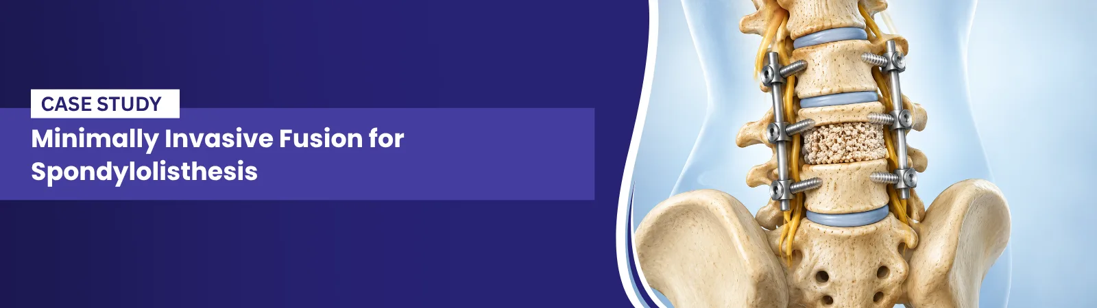
MIS Fusion for Spondylolisthesis
Doctor Profile
Dr. Arvind Umarani
MS Orthopedics, KMC Manipal, Fellowship trained Spine Surgeon
Spine Surgeon | India
Specialisation: Minimally invasive and endoscopic spine surgery, deformity correction, complex spinal fusion
This case study is drawn from published research Dr. Umarani contributed to as part of the spine surgery team at Bombay Hospital and Medical Research Centre, Mumbai. He is credited on the published paper for contributing to the conception and design of the study, and for final approval of the published findings.
Key Takeaways
Condition: High grade spondylolisthesis, meaning severe forward slippage of one vertebra over another, grades III to V
Procedure: Minimally invasive transforaminal lumbar interbody fusion with cantilever reduction, using dome osteotomy and pedicle screw fixation
Doctor: Dr. Arvind Gopalrao Kulkarni, operating surgeon, with Dr. Arvind Umarani as part of the research and clinical team, Bombay Hospital and Medical Research Centre, Mumbai
Technique: Posterior only, single staged approach, mono segmental fusion, no combined anterior surgery required
Outcome: Excellent functional outcome across 36 patients, with significant improvement in back pain, leg pain, and disability scores sustained through 4 years of follow up
Complications: No implant related complications or pseudarthrosis. Two cases of superficial wound infection, both resolved fully with standard treatment
Patient Profile
Field | Group A, Grade III | Group B, Grade IV and V |
Number of patients | 30 | 6 |
Grade of slippage | Grade III | 2 patients Grade IV, 4 patients Grade V |
Level involved | L4 to 5 in 5 patients, L5 to S1 in 25 patients | L4 to 5 in 1 patient, L5 to S1 in 5 patients |
Mean age across full study | 42.16 years | 42.16 years |
Male to female ratio across full study | 1 to 3 | 1 to 3 |
Spinopelvic balance before surgery | Unbalanced in 7 patients | Unbalanced in all patients |
Complete reduction achieved | Included within 24 patients overall | Included within 24 patients overall |
Outcome | Excellent | Excellent |
Patient identities are withheld in line with confidentiality guidelines. The published paper also documents four individual illustrative cases with imaging, ranging from a 15 year old with grade V spondylolisthesis to a 60 year old with grade III spondylolisthesis, each achieving excellent postoperative correction. Data is drawn from Optimising deformity correction research published by Kulkarni AG, Kumar P, Umarani A, Patil S, Chodavadiya S, Asian Spine Journal, 2025, 19(1): 10 to 20.
The Problem
CONDITION
High grade spondylolisthesis means one vertebra has slipped forward over the one below it by more than half its width, most often at the base of the spine where the lumbar spine meets the sacrum. This is a more severe and structurally complex problem than the low grade slippage seen in most spondylolisthesis cases, since it usually comes with a loss of normal spinal curvature, disc collapse, and imbalance in how the pelvis supports the spine. Patients in this study ranged from grade III to grade V, with grade V representing complete displacement of one vertebra off the one beneath it.
EMOTIONAL AND PSYCHOLOGICAL IMPACT
Patients living with high grade spondylolisthesis often carry chronic, disabling low back pain for years before surgery, frequently alongside radiating leg pain that limits walking and daily function. Several patients in this study had already tried years of conservative treatment without relief, and some had visible spinal deformity affecting posture and confidence. For younger patients in particular, including adolescents affected by this condition, the physical limitation and visible change in body alignment can weigh heavily during a stage of life focused on growth and independence.
Consultation And Treatment Plan
WHAT WAS ASSESSED DURING CONSULTATION
Severity and duration of low back pain and radicular leg pain, scored on a standard pain scale
Standing radiographs and whole spine imaging to measure the degree of vertebral slippage
MRI to assess nerve involvement and disc collapse
Most published techniques for grade IV or V spondylolisthesis rely on a combined anterior and posterior surgery, which increases surgical time and recovery. A posterior only approach was chosen wherever technically appropriate to reduce this burden
A cantilever reduction technique was used, where the interbody cage itself acts as a fulcrum, allowing the slipped vertebra to be gradually corrected using the pedicle screws rather than through more aggressive manual manipulation
A dome osteotomy was added in higher grade cases specifically to shorten the affected segment, which protects the exiting nerve root from stretch injury during correction
Continuous neuromonitoring was used throughout every high grade case to track nerve signals in real time during the reduction manoeuvre
This reflects a broader shift many experienced spine surgery teams have made toward less invasive correction, even for structurally severe spinal problems. An Orthopedic Spine Surgeon managing high grade spondylolisthesis today increasingly draws on minimally invasive spine surgery techniques to achieve meaningful correction through a single posterior approach, reserving more extensive combined surgery for cases where it is genuinely required.
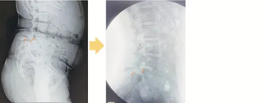
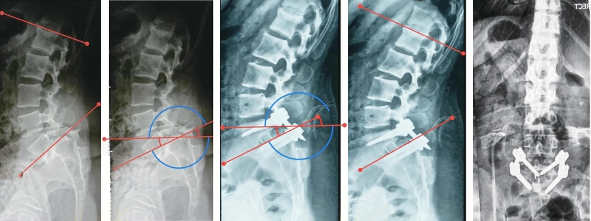
Pre-Operative Photos And Markings
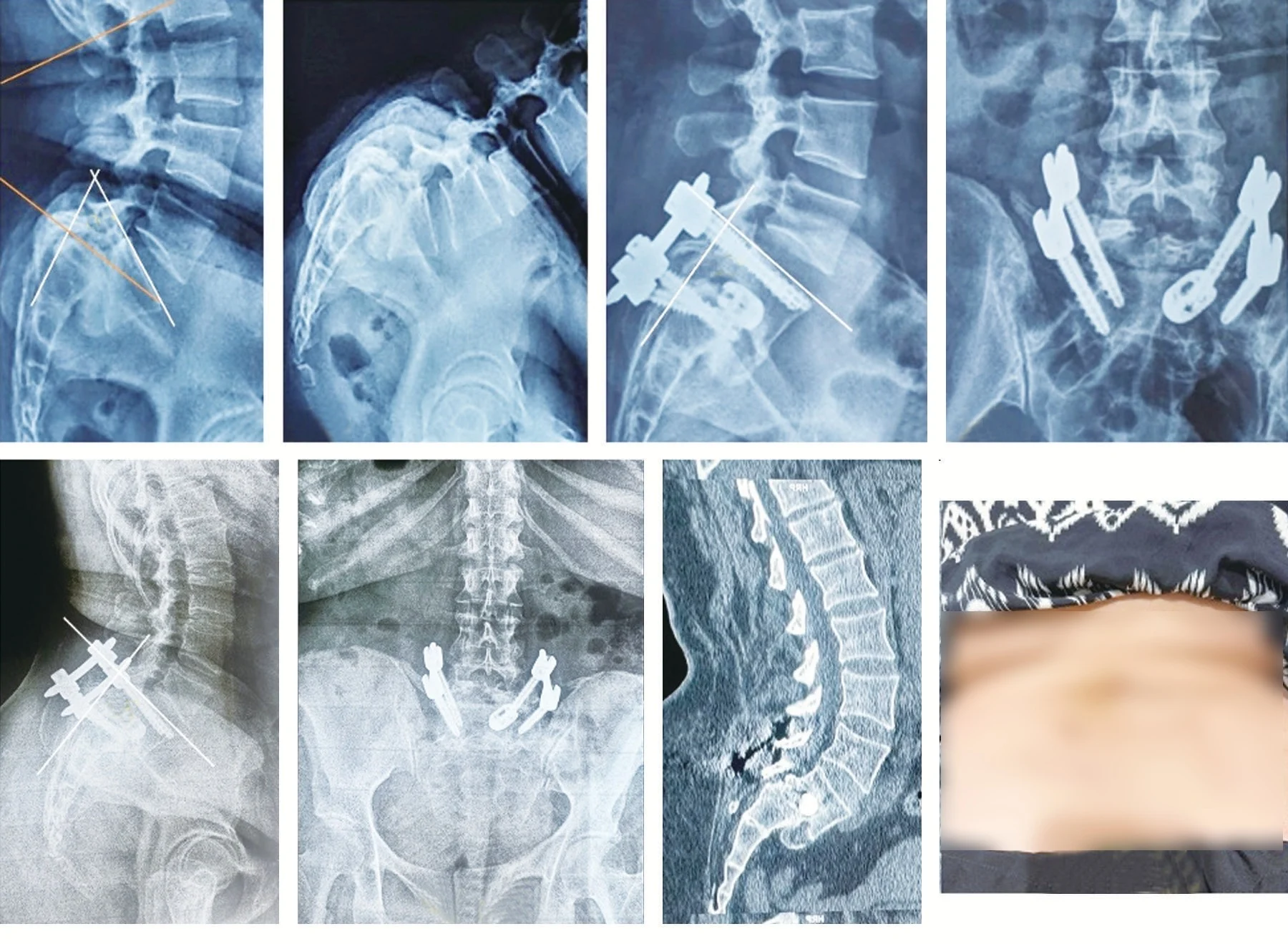
Procedure Details
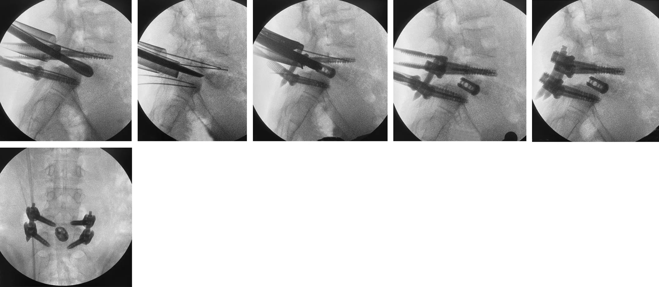
STEP BY STEP OVERVIEW
Patient positioned prone with both hips and knees extended, a specific positioning step used to encourage some correction of the slippage before surgery even begins
Pedicles marked and paramedian incisions made on each side, each approximately 2.5 centimetres, to allow guidewire, screw, and tubular retractor placement
A tubular retractor docked over the affected facet joint, and the relevant bone removed to expose the disc space
A dome osteotomy performed at the sacrum in higher grade cases, shortening the segment to protect the nerve root during correction
Discectomy performed and the disc space prepared, followed by insertion of a bullet shaped cage packed with the patient’s own bone graft
Pedicle screws placed and gradually tightened, using the cage as a fulcrum to achieve reduction of the slipped vertebra
Final position of the screws and cage confirmed, followed by layered closure
Detail | Group A, Grade III | Group B, Grade IV and V |
Approach | Posterior only, single staged surgery | Posterior only, single staged surgery |
Anaesthesia | General anaesthesia | General anaesthesia |
Mean operative time | 225.2 minutes | 270.25 minutes |
Mean blood loss | 140.55 mL | 190.54 mL |
Neuromonitoring | Used for all grade IV and V cases | Used for all patients in this group |
Implant related complications | None | None |
Post-Operative Results
The technique achieved meaningful correction across both patient groups, with complete reduction in the majority of cases and a controlled, partial reduction with restored spinal balance in the remainder. Recovery was steady, with significant improvement in both back pain and leg pain sustained through long term follow up, and no cases of implant failure or failed fusion.
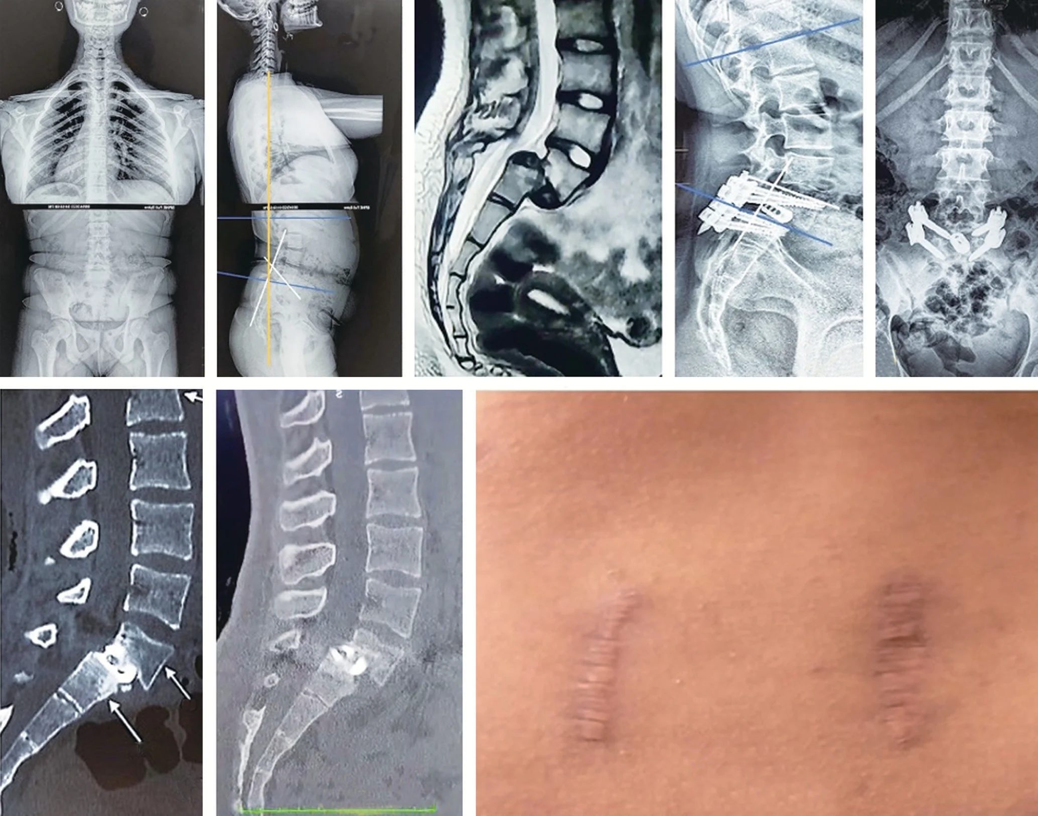
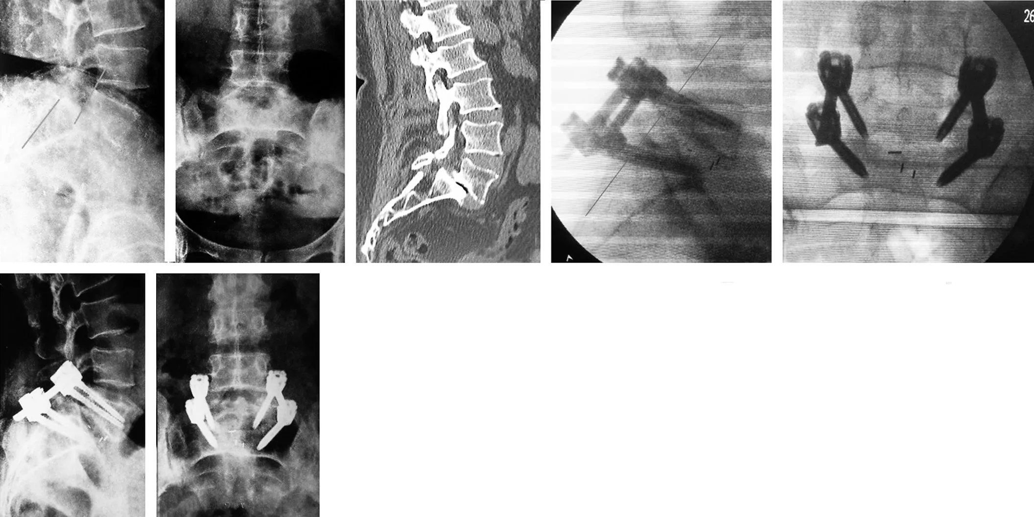
Outcome Metric | Group A, Grade III | Group B, Grade IV and V |
Complete reduction achieved | Included within 24 of 36 total patients | Included within 24 of 36 total patients |
VAS back pain, before surgery | 7.14 out of 10 | 8.0 out of 10 |
VAS back pain, at 4 years | 0.19 out of 10 | 0.29 out of 10 |
VAS leg pain, before surgery | 8.2 out of 10 | 7.3 out of 10 |
VAS leg pain, at 4 years | 0.11 out of 10 | 0.12 out of 10 |
Disability score, before surgery | 25.50 | 28.0 |
Disability score, at 4 years | 5.0 | 5.0 |
Spinopelvic balance restored | Yes, in all patients postoperatively | Yes, in all patients postoperatively |
Neurological injury | None | None |
Patient Feedback
This is a retrospective clinical outcomes analysis, not a marketing testimonial, and it does not include patient quotes in their own words. What the study does confirm directly is that every patient across both groups reported significant, sustained improvement in back pain, leg pain, and disability scores through 4 years of follow up.
Post-Procedure Care And Recovery
INSTRUCTIONS GIVEN TO PATIENTS
Mobilised with a lower spine brace from postoperative Day 1
Discharged on postoperative Day 4
Physiotherapy started the evening of surgery, beginning with gentle ankle and hip exercises
Core strengthening and back extension exercises introduced from 6 weeks postoperatively
Scheduled follow up at 6 weeks, 3 months, 6 months, 1 year, 2 years, 3 years, and 4 years
RECOVERY TIMELINE
Timeframe | What Patient Can Expect |
Day 1 to 4 | Mobilised with a brace, discharged by Day 4 |
6 weeks | Gentle physiotherapy progressing to core strengthening |
6 months | Significant reduction in disability score confirmed |
1 year | Continued improvement in pain and function, fusion assessment underway |
4 years | Sustained pain relief and disability improvement, stable fusion, no loss of correction |
Frequently Asked Questions
Q1. What is high grade spondylolisthesis?
It is a severe form of spinal slippage, where one vertebra has moved forward by more than half its width over the vertebra beneath it. It typically causes chronic low back pain, and often leg pain if a nerve becomes compressed.
Q2. Can severe spinal slippage be corrected without a combined front and back surgery?
In many cases, yes. This study demonstrated that a posterior only, minimally invasive technique achieved excellent correction and long term stability in grade III to V spondylolisthesis, without requiring an additional anterior procedure.
Q3. Is this kind of surgery safe for the nerves during correction?
Continuous nerve monitoring was used throughout every higher grade procedure in this study, and no patient experienced permanent nerve injury or loss of nerve signal during the correction manoeuvre.
Q4. How long does recovery take after this kind of spinal fusion surgery?
Patients were mobilised with a brace from the first day after surgery and discharged within 4 days. Meaningful improvement in pain and disability was already evident by 6 months, with continued improvement sustained through 4 years of follow up.
Considering treatment for severe spinal slippage
If you or a family member has been diagnosed with high grade spondylolisthesis, Dr. Arvind Umarani is glad to walk you through the options available, in plain language and at your own pace.